
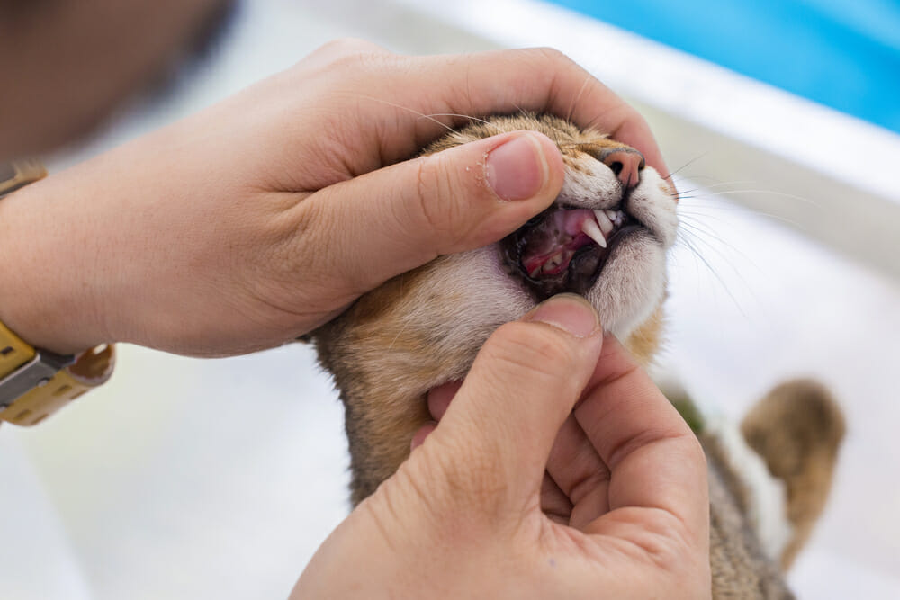
When looking at my own cat’s mouth, I noticed that she had a sore lesion on the gum line that looks like a cavity, I see these frequently in the exam room, and often owners don’t even realize they are there.
These lesions are called feline odontoclastic resorption lesions (FORLs) and are seen in up to 75% of cats over 4 years old. In this disease, cells called odontoclasts, that originate in the bone marrow or spleen, migrate and attach to the external surface of the tooth root often near the gum line and destroy the tooth surface. The odontoclasts are usually involved in the process of turning over deciduous or baby teeth before the permanent teeth erupt. Why these cells remain active in some adult cats is unknown.
With time, the roots of the teeth are completely destroyed, and in the latter stages of the disease, only the portion of the tooth above the gum line is visible, and it may also be affected. In many cats, the end-stage affected tooth is missing.
FORLs are not a cavity, although they have incorrectly been called that. Bacteria cause cavities, and although the true cause of FORLs is unknown, they are not a bacterial disease. There is a significant amount of research in the veterinary dentistry field as to the cause of FORLs.
Cats with FORLs are often identified as having oral pain, salivating and having difficulty eating. When we examine these cats, we will find missing teeth or teeth that have portions of the tooth above the gum line (crown) missing. In areas where portions of the crown are missing, the gums will often fill into the lesion, and a red spot is noticed on the tooth. The teeth with early FORLs cannot always be identified on clinical examination, because the disease can be localized only to the root surface below the gum line and it can only be observed on radiographs (x-rays). Symptomatic cats are usually cats that have teeth with partially missing tooth crowns, and where the disease process has moved beyond the root surface, with these cats even touching that tooth can cause significant chattering due to oral discomfort. My cat didn’t show any obvious clinical signs but did chatter when I touched the tooth with my finger, so I knew that it was sore.
Since only the end-stage lesions involving the tooth crown can be identified readily on clinical examination, the remainder of lesions must be diagnosed by dental radiographs. Due to a high percentage of cats affected by this disease, cats over the age of 4 are recommended to have dental radiographs as a screening test for the disease when having their teeth cleaned.
FORLs are a painful disease in the cat, and cats with this disease should be treated. The primary treatment for this disease is extraction or crown amputation of the affected teeth. Crown amputation is a procedure where the crown of the affected tooth is removed, leaving the resorbing roots buried in the bone to continue resorbing on their own. The crown amputation procedure alleviates the clinical signs of disease because the exposed and sensitive portion of the tooth is removed. This procedure is only performed on affected teeth that have been appropriately radiographed and have severe tooth root resorption.
The prognosis following extraction or crown amputation of affected teeth is good. Still, affected cats will always have a predisposition to the development of additional lesions in their remaining teeth, and should be checked annually.
In my own cat Mia, I took dental radiographs and saw that the tooth with the obvious lesion had a resorbing root and I performed a crown amputation, the same premolar on the other side of her mouth was also affected when looking at the radiographs even though it wasn’t clinically visible, I did a crown amputation on that tooth as well and Mia recovered uneventfully.
Some information collected from VetMedtext, author: Anson J. Tsugawa
Written by Jane Corkum, DVM